Neuromuscular Junction in Nervous System when Doing Sports. All the activities of the human body conscious or unconscious, which is coordinated by the nervous system in collaboration with the hormone system as the central regulator. The nervous system consists of long threads which extend from the brain, spinal cord and ganglion which spread throughout the body. Activities such as typing and writing is coordinated by the nervous system in the form of coordinate eyes with hand, as well as sports activities such as running, swimming, etc., coordinated by the nervous system.
When the nervous system giving orders to the muscles then there was a movement. The order propagated through nerve cells in the body to muscles, the nervous system to the muscle fibers, there is such a junction that caused the movement called the neuromuscular junction.
At the time of doing physical activity or sports our bodies move according to orders of the nervous system. The order was delivered to the muscles through the neuromuscular junction, nerve impulses are delivered with the help of neurotransmitter fluid. The application of the concept of the neuromuscular junction in sports activities and the occurrence of muscle movements that originated from the stimulation, then the nerve giving commands to the muscles through the neuromuscular junction as well as the effect of the intensity of an excessive activity of the neuromuscular junction.
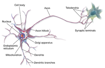
A. Anatomy of Nerve Cells (Neuron)

- The cell body
- Neurites (axons) is the tail of the nerve cells.
- Dendrite is part of the cell body and serves to receive a stimulus.
B. The type of nerves with its function
1. The Motor Nerves
Its function: to bring the stimulation of the central nervous system heading to muscles and glands, as a result is muscles tighten (contraction) and glands secrete sap.
2. The Sensory Nerves
Its functions: nerves that carry stimuli from outside the body to the central nervous.
Its function: to bring the stimulation of the central nervous system heading to muscles and glands, as a result is muscles tighten (contraction) and glands secrete sap.
2. The Sensory Nerves
Its functions: nerves that carry stimuli from outside the body to the central nervous.
C. Synapse
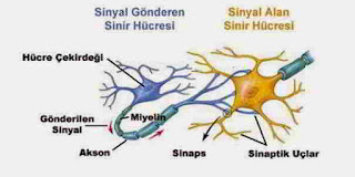
 Synapses are the points of contact between the axon terminal of one neuron to another neuron. Synapses are formed by terminal axons swell. In the cytoplasm of synapses, there are synaptic vesicles. When the impulse reaches the end of the neuron, the vesicles would move, and then fused with pre-synaptic membrane and release acetylcholine. Acetylcholine diffuses through the synaptic gap, and then attaches to receptors on the post-synaptic membrane. Adherence acetylcholine receptor causing subsequent impulses in nerve cells. The enzyme acetylcholinesterase, outlining acetylcholine whose task has been completed.
D. Nerve Impulses
In the event of propagation of nerve impulses, there is medium that play an important role, namely the membrane potential. Membrane potential is a membrane capable of delivering impulses and make changes in electrical potential along both sides of the membrane.
The term with impulse propagation events:
The term with impulse propagation events:
- Polarization membranes: the membranes of nerve cells in normal circumstances.
- Depolarization of the membrane: the cell membrane of nerve excited / happening electrical currents propagated along the membrane.
- Repolarization of the membrane: the recovery process a positive charge and a negative charge is present in the membrane of nerve cells in normal circumstances
E. Neuromuscular Junction
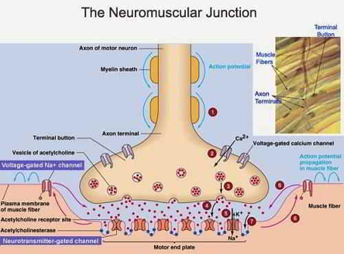
A nerve bundles have branches that control some of the muscle fibers. Approximately 100-150 muscle fibers can be controlled by one motor unit (motor nerves along the muscle fibers it serves). The relationship between the nerve fibers resulting in a movement called the neuromuscular junction or myoneural junction. For more details can be seen in the image below.
 In the relationship between nerve to muscles, nerve fibers edge was released a fluids when there is a signal. Fluid that comes out from edge of nerve called: chemical transmitter, eg acetyl choline. Acetyl choline serves as a substance that is able to excite muscle fibers resulting in an electric current and increase the permeability of the membrane so that the muscle membrane easy to follow / covered with impulse. With these circumstances will accelerate the movement by effector.

F. The Mechanism of Muscle Contraction
The mechanism of Muscle Contraction start with action potential (action potential is positive and negative ionic flow moving in the cell membrane) in fibers - muscle fibers. This gives rise to action potential electrical currents that spread to the inside of the fibers, which causes the release of calcium ions from the sarcoplasmic reticulum. Furthermore, the calcium ions cause chemical events contraction process.
G. Muscle fibers by Nerve Stimulation
In the normal body functions, muscle fibers are stimulated by myelinated nerve fibers. Nerve fibers attached to the muscle fibers in a muscles nerve connections (neuromuscular junction) located in the middle of muscles. When the action potential until at the neuromuscular junction, happened depolarization of the neuronal membrane, causing Acethylcholin released, then will be attached to the motor end plate membrane, causing the release of calcium ions which cause bond actin - myosin ultimately happened muscle contraction. Therefore, the action potential fibers spread from the middle towards both ends, so that contraction occurred almost simultaneously throughout muscles sarcomere.
Movements caused by muscle contraction appears from their command in the form of nerve impulses that are sent through the nerve cells that exist in our bodies. The propagation impulse is delivered to muscles through the neuromuscular junction so it came to what was ordered by the nerves to the muscles.
Thats the article about neuromuscular junction in nervous system when doing sports. May be useful for the readers, do not forget to look at other interesting articles on this website. Thank you.
